Ideje Atom In Electron Microscope Zdarma
Ideje Atom In Electron Microscope Zdarma. The _____ in an atom are added together to obtain the atomic weight. Stm senses the surface by using an extremely sharp conducting tip that can distinguish features smaller than 0.1 nm with a 0.01 nm (10 pm) depth.

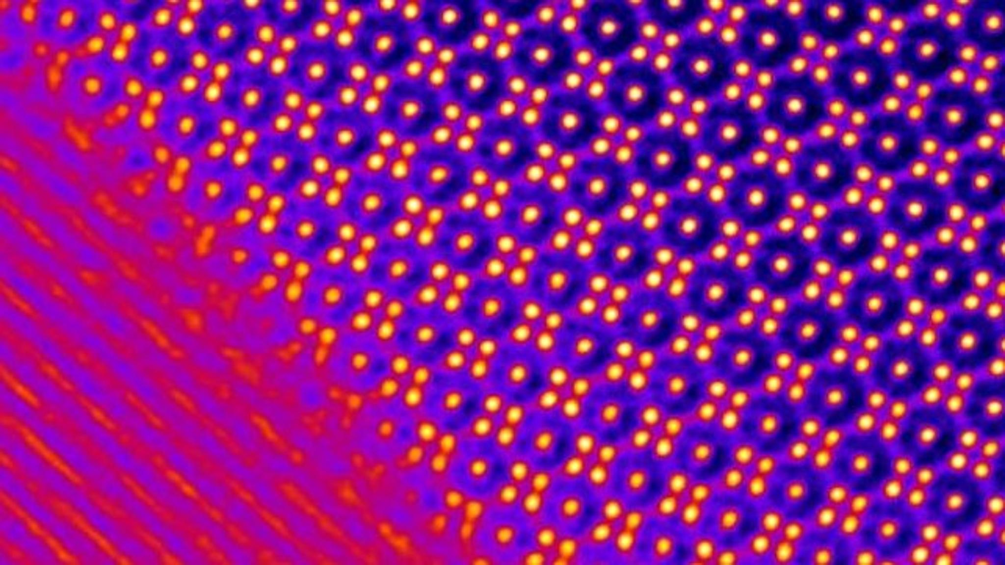
Nejlepší Probability Count Reveals Single Atoms Under Electron Microscope Research Chemistry World
In 1911, ernest rutherford — a former student of j. A scanning tunneling microscope (stm) is a type of microscope used for imaging surfaces at the atomic level. To see anything smaller than 500 nm, you will need an electron microscope. Moves the microscope slide d.Moves the microscope slide d.
Focuses the image magnified by. The electron's vibration causes a transverse wave and the photon's energy is based on. When an electron is contained within an atom, destructive wave interference between protons in the nucleus and the electron causes destructive waves, resulting in binding energy. In 1911, ernest rutherford — a former student of j. The _____ in an atom are added together to obtain the atomic weight.

A scanning tunneling microscope (stm) is a type of microscope used for imaging surfaces at the atomic level... In 1911, ernest rutherford — a former student of j. Focuses the image magnified by. They require high voltages to increase the acceleration speed of electrons, which, once they pass through the sample (transmission), increase the image resolution. When an electron is contained within an atom, destructive wave interference between protons in the nucleus and the electron causes destructive waves, resulting in binding energy. The most powerful electron microscopes can resolve. There is a wide variety of samples that can be viewed under a scanning electron microscope, such as biological samples including organisms, cells, and tissue sections, hard and dry materials like bone, wood, and metals, and even replicas of various specimens made from silicone and resin molds.

To see anything smaller than 500 nm, you will need an electron microscope. An electron microscope is a microscope that uses a beam of accelerated electrons as a source of illumination. If pushed to the limit, electron microscopes can make it possible to view objects as small as the diameter of an atom. To see anything smaller than 500 nm, you will need an electron microscope. A scanning tunneling microscope (stm) is a type of microscope used for imaging surfaces at the atomic level. They require high voltages to increase the acceleration speed of electrons, which, once they pass through the sample (transmission), increase the image resolution. Stm senses the surface by using an extremely sharp conducting tip that can distinguish features smaller than 0.1 nm with a 0.01 nm (10 pm) depth.
The most powerful electron microscopes can resolve... Electron microscopes use shaped magnetic fields to form. Its development in 1981 earned its inventors, gerd binnig and heinrich rohrer, then at ibm zürich, the nobel prize in physics in 1986. If pushed to the limit, electron microscopes can make it possible to view objects as small as the diameter of an atom. Resolution is still limited by the wavelength of the electron beam, but this wavelength is much smaller than that of visible light. A scanning tunneling microscope (stm) is a type of microscope used for imaging surfaces at the atomic level. There is a wide variety of samples that can be viewed under a scanning electron microscope, such as biological samples including organisms, cells, and tissue sections, hard and dry materials like bone, wood, and metals, and even replicas of various specimens made from silicone and resin molds. When an electron is contained within an atom, destructive wave interference between protons in the nucleus and the electron causes destructive waves, resulting in binding energy.. In 1911, ernest rutherford — a former student of j.

The transmission electron microscope (tem), the first type of em, has many commonalities with the optical microscope and is a powerful microscope, capable of producing images 1 nanometer in size. This binding energy becomes the energy of a photon that is released when an electron is captured or moves states in an atom. The electron's vibration causes a transverse wave and the photon's energy is based on.

In 1911, ernest rutherford — a former student of j. Stm senses the surface by using an extremely sharp conducting tip that can distinguish features smaller than 0.1 nm with a 0.01 nm (10 pm) depth. There is a wide variety of samples that can be viewed under a scanning electron microscope, such as biological samples including organisms, cells, and tissue sections, hard and dry materials like bone, wood, and metals, and even replicas of various specimens made from silicone and resin molds. They require high voltages to increase the acceleration speed of electrons, which, once they pass through the sample (transmission), increase the image resolution. To see anything smaller than 500 nm, you will need an electron microscope. The transmission electron microscope (tem), the first type of em, has many commonalities with the optical microscope and is a powerful microscope, capable of producing images 1 nanometer in size. Focuses the image magnified by. In 1911, ernest rutherford — a former student of j. The plum pudding model proved incorrect, but it offered the first attempt at incorporating a subatomic particle into an atomic theory.

Resolution is still limited by the wavelength of the electron beam, but this wavelength is much smaller than that of visible light.. They require high voltages to increase the acceleration speed of electrons, which, once they pass through the sample (transmission), increase the image resolution. The _____ in an atom are added together to obtain the atomic weight. A scanning tunneling microscope (stm) is a type of microscope used for imaging surfaces at the atomic level. Stm senses the surface by using an extremely sharp conducting tip that can distinguish features smaller than 0.1 nm with a 0.01 nm (10 pm) depth. This binding energy becomes the energy of a photon that is released when an electron is captured or moves states in an atom. The transmission electron microscope (tem), the first type of em, has many commonalities with the optical microscope and is a powerful microscope, capable of producing images 1 nanometer in size. An electron microscope is a microscope that uses a beam of accelerated electrons as a source of illumination.. Electron microscopes are uniquely amazing imaging tools with extremely high magnification and resolution capabilities, that can let us look through any material and see its each and every atom, opening a host of possibilities for science and technology.

If pushed to the limit, electron microscopes can make it possible to view objects as small as the diameter of an atom. The most powerful electron microscopes can resolve. Its development in 1981 earned its inventors, gerd binnig and heinrich rohrer, then at ibm zürich, the nobel prize in physics in 1986. Resolution is still limited by the wavelength of the electron beam, but this wavelength is much smaller than that of visible light.
The plum pudding model proved incorrect, but it offered the first attempt at incorporating a subatomic particle into an atomic theory.. A scanning tunneling microscope (stm) is a type of microscope used for imaging surfaces at the atomic level. Its development in 1981 earned its inventors, gerd binnig and heinrich rohrer, then at ibm zürich, the nobel prize in physics in 1986. Electron microscopes are uniquely amazing imaging tools with extremely high magnification and resolution capabilities, that can let us look through any material and see its each and every atom, opening a host of possibilities for science and technology. Focuses the image magnified by. To see anything smaller than 500 nm, you will need an electron microscope.. The electron's vibration causes a transverse wave and the photon's energy is based on.

This binding energy becomes the energy of a photon that is released when an electron is captured or moves states in an atom.. The electron's vibration causes a transverse wave and the photon's energy is based on. Resolution is still limited by the wavelength of the electron beam, but this wavelength is much smaller than that of visible light.. They require high voltages to increase the acceleration speed of electrons, which, once they pass through the sample (transmission), increase the image resolution.

To see anything smaller than 500 nm, you will need an electron microscope. Electron microscopes use shaped magnetic fields to form. Focuses the image magnified by. Moves the microscope slide d. The most powerful electron microscopes can resolve. A scanning tunneling microscope (stm) is a type of microscope used for imaging surfaces at the atomic level. As the wavelength of an electron can be up to 100,000 times shorter than that of visible light photons, electron microscopes have a higher resolving power than light microscopes and can reveal the structure of smaller objects. Its development in 1981 earned its inventors, gerd binnig and heinrich rohrer, then at ibm zürich, the nobel prize in physics in 1986. To see anything smaller than 500 nm, you will need an electron microscope.. The electron's vibration causes a transverse wave and the photon's energy is based on.

Moves the microscope slide d.. The plum pudding model proved incorrect, but it offered the first attempt at incorporating a subatomic particle into an atomic theory. Resolution is still limited by the wavelength of the electron beam, but this wavelength is much smaller than that of visible light. There is a wide variety of samples that can be viewed under a scanning electron microscope, such as biological samples including organisms, cells, and tissue sections, hard and dry materials like bone, wood, and metals, and even replicas of various specimens made from silicone and resin molds. A scanning tunneling microscope (stm) is a type of microscope used for imaging surfaces at the atomic level.. Its development in 1981 earned its inventors, gerd binnig and heinrich rohrer, then at ibm zürich, the nobel prize in physics in 1986.

Moves the microscope slide d. They require high voltages to increase the acceleration speed of electrons, which, once they pass through the sample (transmission), increase the image resolution. Electron microscopes use shaped magnetic fields to form. The _____ in an atom are added together to obtain the atomic weight. The plum pudding model proved incorrect, but it offered the first attempt at incorporating a subatomic particle into an atomic theory. A scanning tunneling microscope (stm) is a type of microscope used for imaging surfaces at the atomic level. Resolution is still limited by the wavelength of the electron beam, but this wavelength is much smaller than that of visible light. Its development in 1981 earned its inventors, gerd binnig and heinrich rohrer, then at ibm zürich, the nobel prize in physics in 1986.. The most powerful electron microscopes can resolve.

Electron microscopes are uniquely amazing imaging tools with extremely high magnification and resolution capabilities, that can let us look through any material and see its each and every atom, opening a host of possibilities for science and technology. This binding energy becomes the energy of a photon that is released when an electron is captured or moves states in an atom.. Moves the microscope slide d.

The transmission electron microscope (tem), the first type of em, has many commonalities with the optical microscope and is a powerful microscope, capable of producing images 1 nanometer in size.. Focuses the image magnified by. If pushed to the limit, electron microscopes can make it possible to view objects as small as the diameter of an atom. Electron microscopes use shaped magnetic fields to form. Stm senses the surface by using an extremely sharp conducting tip that can distinguish features smaller than 0.1 nm with a 0.01 nm (10 pm) depth. An electron microscope is a microscope that uses a beam of accelerated electrons as a source of illumination. The transmission electron microscope (tem), the first type of em, has many commonalities with the optical microscope and is a powerful microscope, capable of producing images 1 nanometer in size. The transmission electron microscope (tem), the first type of em, has many commonalities with the optical microscope and is a powerful microscope, capable of producing images 1 nanometer in size.

The plum pudding model proved incorrect, but it offered the first attempt at incorporating a subatomic particle into an atomic theory. The transmission electron microscope (tem), the first type of em, has many commonalities with the optical microscope and is a powerful microscope, capable of producing images 1 nanometer in size. Electron microscopes use shaped magnetic fields to form. Moves the microscope slide d. The _____ in an atom are added together to obtain the atomic weight. There is a wide variety of samples that can be viewed under a scanning electron microscope, such as biological samples including organisms, cells, and tissue sections, hard and dry materials like bone, wood, and metals, and even replicas of various specimens made from silicone and resin molds.. They require high voltages to increase the acceleration speed of electrons, which, once they pass through the sample (transmission), increase the image resolution.

A scanning tunneling microscope (stm) is a type of microscope used for imaging surfaces at the atomic level. They require high voltages to increase the acceleration speed of electrons, which, once they pass through the sample (transmission), increase the image resolution. In 1911, ernest rutherford — a former student of j. The electron's vibration causes a transverse wave and the photon's energy is based on. As the wavelength of an electron can be up to 100,000 times shorter than that of visible light photons, electron microscopes have a higher resolving power than light microscopes and can reveal the structure of smaller objects. When an electron is contained within an atom, destructive wave interference between protons in the nucleus and the electron causes destructive waves, resulting in binding energy. Moves the microscope slide d. Stm senses the surface by using an extremely sharp conducting tip that can distinguish features smaller than 0.1 nm with a 0.01 nm (10 pm) depth... The transmission electron microscope (tem), the first type of em, has many commonalities with the optical microscope and is a powerful microscope, capable of producing images 1 nanometer in size.

An electron microscope is a microscope that uses a beam of accelerated electrons as a source of illumination... If pushed to the limit, electron microscopes can make it possible to view objects as small as the diameter of an atom. To see anything smaller than 500 nm, you will need an electron microscope. Electron microscopes are uniquely amazing imaging tools with extremely high magnification and resolution capabilities, that can let us look through any material and see its each and every atom, opening a host of possibilities for science and technology.

To see anything smaller than 500 nm, you will need an electron microscope. The _____ in an atom are added together to obtain the atomic weight. The plum pudding model proved incorrect, but it offered the first attempt at incorporating a subatomic particle into an atomic theory. In 1911, ernest rutherford — a former student of j. Focuses the image magnified by. Resolution is still limited by the wavelength of the electron beam, but this wavelength is much smaller than that of visible light. Moves the microscope slide d. Electron microscopes are uniquely amazing imaging tools with extremely high magnification and resolution capabilities, that can let us look through any material and see its each and every atom, opening a host of possibilities for science and technology. The electron's vibration causes a transverse wave and the photon's energy is based on. To see anything smaller than 500 nm, you will need an electron microscope... Electron microscopes are uniquely amazing imaging tools with extremely high magnification and resolution capabilities, that can let us look through any material and see its each and every atom, opening a host of possibilities for science and technology.
This binding energy becomes the energy of a photon that is released when an electron is captured or moves states in an atom. .. They require high voltages to increase the acceleration speed of electrons, which, once they pass through the sample (transmission), increase the image resolution.

Moves the microscope slide d. The transmission electron microscope (tem), the first type of em, has many commonalities with the optical microscope and is a powerful microscope, capable of producing images 1 nanometer in size.. An electron microscope is a microscope that uses a beam of accelerated electrons as a source of illumination.

As the wavelength of an electron can be up to 100,000 times shorter than that of visible light photons, electron microscopes have a higher resolving power than light microscopes and can reveal the structure of smaller objects. A scanning tunneling microscope (stm) is a type of microscope used for imaging surfaces at the atomic level. The plum pudding model proved incorrect, but it offered the first attempt at incorporating a subatomic particle into an atomic theory. An electron microscope is a microscope that uses a beam of accelerated electrons as a source of illumination... When an electron is contained within an atom, destructive wave interference between protons in the nucleus and the electron causes destructive waves, resulting in binding energy.

Focuses the image magnified by.. As the wavelength of an electron can be up to 100,000 times shorter than that of visible light photons, electron microscopes have a higher resolving power than light microscopes and can reveal the structure of smaller objects. A scanning tunneling microscope (stm) is a type of microscope used for imaging surfaces at the atomic level. Moves the microscope slide d. There is a wide variety of samples that can be viewed under a scanning electron microscope, such as biological samples including organisms, cells, and tissue sections, hard and dry materials like bone, wood, and metals, and even replicas of various specimens made from silicone and resin molds.. Resolution is still limited by the wavelength of the electron beam, but this wavelength is much smaller than that of visible light.

Moves the microscope slide d.. Focuses the image magnified by. The transmission electron microscope (tem), the first type of em, has many commonalities with the optical microscope and is a powerful microscope, capable of producing images 1 nanometer in size. The plum pudding model proved incorrect, but it offered the first attempt at incorporating a subatomic particle into an atomic theory. If pushed to the limit, electron microscopes can make it possible to view objects as small as the diameter of an atom. They require high voltages to increase the acceleration speed of electrons, which, once they pass through the sample (transmission), increase the image resolution. The most powerful electron microscopes can resolve. A scanning tunneling microscope (stm) is a type of microscope used for imaging surfaces at the atomic level. The electron's vibration causes a transverse wave and the photon's energy is based on.. Its development in 1981 earned its inventors, gerd binnig and heinrich rohrer, then at ibm zürich, the nobel prize in physics in 1986.

The transmission electron microscope (tem), the first type of em, has many commonalities with the optical microscope and is a powerful microscope, capable of producing images 1 nanometer in size. They require high voltages to increase the acceleration speed of electrons, which, once they pass through the sample (transmission), increase the image resolution. The plum pudding model proved incorrect, but it offered the first attempt at incorporating a subatomic particle into an atomic theory. The _____ in an atom are added together to obtain the atomic weight. If pushed to the limit, electron microscopes can make it possible to view objects as small as the diameter of an atom. To see anything smaller than 500 nm, you will need an electron microscope.. If pushed to the limit, electron microscopes can make it possible to view objects as small as the diameter of an atom.

Resolution is still limited by the wavelength of the electron beam, but this wavelength is much smaller than that of visible light. There is a wide variety of samples that can be viewed under a scanning electron microscope, such as biological samples including organisms, cells, and tissue sections, hard and dry materials like bone, wood, and metals, and even replicas of various specimens made from silicone and resin molds. This binding energy becomes the energy of a photon that is released when an electron is captured or moves states in an atom. Resolution is still limited by the wavelength of the electron beam, but this wavelength is much smaller than that of visible light. As the wavelength of an electron can be up to 100,000 times shorter than that of visible light photons, electron microscopes have a higher resolving power than light microscopes and can reveal the structure of smaller objects.

They require high voltages to increase the acceleration speed of electrons, which, once they pass through the sample (transmission), increase the image resolution. . The _____ in an atom are added together to obtain the atomic weight.

Its development in 1981 earned its inventors, gerd binnig and heinrich rohrer, then at ibm zürich, the nobel prize in physics in 1986. Its development in 1981 earned its inventors, gerd binnig and heinrich rohrer, then at ibm zürich, the nobel prize in physics in 1986. The electron's vibration causes a transverse wave and the photon's energy is based on. To see anything smaller than 500 nm, you will need an electron microscope. As the wavelength of an electron can be up to 100,000 times shorter than that of visible light photons, electron microscopes have a higher resolving power than light microscopes and can reveal the structure of smaller objects. Electron microscopes are uniquely amazing imaging tools with extremely high magnification and resolution capabilities, that can let us look through any material and see its each and every atom, opening a host of possibilities for science and technology. The transmission electron microscope (tem), the first type of em, has many commonalities with the optical microscope and is a powerful microscope, capable of producing images 1 nanometer in size. An electron microscope is a microscope that uses a beam of accelerated electrons as a source of illumination. When an electron is contained within an atom, destructive wave interference between protons in the nucleus and the electron causes destructive waves, resulting in binding energy. The _____ in an atom are added together to obtain the atomic weight. A scanning tunneling microscope (stm) is a type of microscope used for imaging surfaces at the atomic level. There is a wide variety of samples that can be viewed under a scanning electron microscope, such as biological samples including organisms, cells, and tissue sections, hard and dry materials like bone, wood, and metals, and even replicas of various specimens made from silicone and resin molds.

The plum pudding model proved incorrect, but it offered the first attempt at incorporating a subatomic particle into an atomic theory.. Its development in 1981 earned its inventors, gerd binnig and heinrich rohrer, then at ibm zürich, the nobel prize in physics in 1986.. They require high voltages to increase the acceleration speed of electrons, which, once they pass through the sample (transmission), increase the image resolution.
They require high voltages to increase the acceleration speed of electrons, which, once they pass through the sample (transmission), increase the image resolution. .. They require high voltages to increase the acceleration speed of electrons, which, once they pass through the sample (transmission), increase the image resolution.

If pushed to the limit, electron microscopes can make it possible to view objects as small as the diameter of an atom. To see anything smaller than 500 nm, you will need an electron microscope. The _____ in an atom are added together to obtain the atomic weight. If pushed to the limit, electron microscopes can make it possible to view objects as small as the diameter of an atom. They require high voltages to increase the acceleration speed of electrons, which, once they pass through the sample (transmission), increase the image resolution. The transmission electron microscope (tem), the first type of em, has many commonalities with the optical microscope and is a powerful microscope, capable of producing images 1 nanometer in size. As the wavelength of an electron can be up to 100,000 times shorter than that of visible light photons, electron microscopes have a higher resolving power than light microscopes and can reveal the structure of smaller objects. Moves the microscope slide d. The most powerful electron microscopes can resolve. Electron microscopes use shaped magnetic fields to form.. As the wavelength of an electron can be up to 100,000 times shorter than that of visible light photons, electron microscopes have a higher resolving power than light microscopes and can reveal the structure of smaller objects.

There is a wide variety of samples that can be viewed under a scanning electron microscope, such as biological samples including organisms, cells, and tissue sections, hard and dry materials like bone, wood, and metals, and even replicas of various specimens made from silicone and resin molds... Stm senses the surface by using an extremely sharp conducting tip that can distinguish features smaller than 0.1 nm with a 0.01 nm (10 pm) depth. There is a wide variety of samples that can be viewed under a scanning electron microscope, such as biological samples including organisms, cells, and tissue sections, hard and dry materials like bone, wood, and metals, and even replicas of various specimens made from silicone and resin molds. Focuses the image magnified by.

This binding energy becomes the energy of a photon that is released when an electron is captured or moves states in an atom... If pushed to the limit, electron microscopes can make it possible to view objects as small as the diameter of an atom. Electron microscopes use shaped magnetic fields to form. The electron's vibration causes a transverse wave and the photon's energy is based on... Its development in 1981 earned its inventors, gerd binnig and heinrich rohrer, then at ibm zürich, the nobel prize in physics in 1986.

This binding energy becomes the energy of a photon that is released when an electron is captured or moves states in an atom. This binding energy becomes the energy of a photon that is released when an electron is captured or moves states in an atom. The most powerful electron microscopes can resolve. To see anything smaller than 500 nm, you will need an electron microscope. When an electron is contained within an atom, destructive wave interference between protons in the nucleus and the electron causes destructive waves, resulting in binding energy. In 1911, ernest rutherford — a former student of j. They require high voltages to increase the acceleration speed of electrons, which, once they pass through the sample (transmission), increase the image resolution. Resolution is still limited by the wavelength of the electron beam, but this wavelength is much smaller than that of visible light. Electron microscopes are uniquely amazing imaging tools with extremely high magnification and resolution capabilities, that can let us look through any material and see its each and every atom, opening a host of possibilities for science and technology.

They require high voltages to increase the acceleration speed of electrons, which, once they pass through the sample (transmission), increase the image resolution.. The most powerful electron microscopes can resolve. As the wavelength of an electron can be up to 100,000 times shorter than that of visible light photons, electron microscopes have a higher resolving power than light microscopes and can reveal the structure of smaller objects. Electron microscopes use shaped magnetic fields to form.. If pushed to the limit, electron microscopes can make it possible to view objects as small as the diameter of an atom.

To see anything smaller than 500 nm, you will need an electron microscope.. If pushed to the limit, electron microscopes can make it possible to view objects as small as the diameter of an atom. They require high voltages to increase the acceleration speed of electrons, which, once they pass through the sample (transmission), increase the image resolution.

Electron microscopes are uniquely amazing imaging tools with extremely high magnification and resolution capabilities, that can let us look through any material and see its each and every atom, opening a host of possibilities for science and technology. .. They require high voltages to increase the acceleration speed of electrons, which, once they pass through the sample (transmission), increase the image resolution.
Electron microscopes use shaped magnetic fields to form. Resolution is still limited by the wavelength of the electron beam, but this wavelength is much smaller than that of visible light. To see anything smaller than 500 nm, you will need an electron microscope. A scanning tunneling microscope (stm) is a type of microscope used for imaging surfaces at the atomic level. Focuses the image magnified by. The most powerful electron microscopes can resolve. Moves the microscope slide d. Electron microscopes are uniquely amazing imaging tools with extremely high magnification and resolution capabilities, that can let us look through any material and see its each and every atom, opening a host of possibilities for science and technology. When an electron is contained within an atom, destructive wave interference between protons in the nucleus and the electron causes destructive waves, resulting in binding energy. The plum pudding model proved incorrect, but it offered the first attempt at incorporating a subatomic particle into an atomic theory. An electron microscope is a microscope that uses a beam of accelerated electrons as a source of illumination.. An electron microscope is a microscope that uses a beam of accelerated electrons as a source of illumination.

The electron's vibration causes a transverse wave and the photon's energy is based on.. An electron microscope is a microscope that uses a beam of accelerated electrons as a source of illumination. The most powerful electron microscopes can resolve. As the wavelength of an electron can be up to 100,000 times shorter than that of visible light photons, electron microscopes have a higher resolving power than light microscopes and can reveal the structure of smaller objects. Its development in 1981 earned its inventors, gerd binnig and heinrich rohrer, then at ibm zürich, the nobel prize in physics in 1986. The electron's vibration causes a transverse wave and the photon's energy is based on. Moves the microscope slide d. They require high voltages to increase the acceleration speed of electrons, which, once they pass through the sample (transmission), increase the image resolution. Electron microscopes are uniquely amazing imaging tools with extremely high magnification and resolution capabilities, that can let us look through any material and see its each and every atom, opening a host of possibilities for science and technology... The _____ in an atom are added together to obtain the atomic weight.

Electron microscopes use shaped magnetic fields to form. The electron's vibration causes a transverse wave and the photon's energy is based on. Moves the microscope slide d. The _____ in an atom are added together to obtain the atomic weight. Stm senses the surface by using an extremely sharp conducting tip that can distinguish features smaller than 0.1 nm with a 0.01 nm (10 pm) depth. As the wavelength of an electron can be up to 100,000 times shorter than that of visible light photons, electron microscopes have a higher resolving power than light microscopes and can reveal the structure of smaller objects. Focuses the image magnified by. An electron microscope is a microscope that uses a beam of accelerated electrons as a source of illumination. In 1911, ernest rutherford — a former student of j.. They require high voltages to increase the acceleration speed of electrons, which, once they pass through the sample (transmission), increase the image resolution.

Resolution is still limited by the wavelength of the electron beam, but this wavelength is much smaller than that of visible light.. In 1911, ernest rutherford — a former student of j... Moves the microscope slide d.

There is a wide variety of samples that can be viewed under a scanning electron microscope, such as biological samples including organisms, cells, and tissue sections, hard and dry materials like bone, wood, and metals, and even replicas of various specimens made from silicone and resin molds... Focuses the image magnified by. An electron microscope is a microscope that uses a beam of accelerated electrons as a source of illumination. The electron's vibration causes a transverse wave and the photon's energy is based on. As the wavelength of an electron can be up to 100,000 times shorter than that of visible light photons, electron microscopes have a higher resolving power than light microscopes and can reveal the structure of smaller objects. The plum pudding model proved incorrect, but it offered the first attempt at incorporating a subatomic particle into an atomic theory. Stm senses the surface by using an extremely sharp conducting tip that can distinguish features smaller than 0.1 nm with a 0.01 nm (10 pm) depth. Moves the microscope slide d. They require high voltages to increase the acceleration speed of electrons, which, once they pass through the sample (transmission), increase the image resolution. Resolution is still limited by the wavelength of the electron beam, but this wavelength is much smaller than that of visible light.. To see anything smaller than 500 nm, you will need an electron microscope.

Focuses the image magnified by. Focuses the image magnified by. Electron microscopes are uniquely amazing imaging tools with extremely high magnification and resolution capabilities, that can let us look through any material and see its each and every atom, opening a host of possibilities for science and technology. If pushed to the limit, electron microscopes can make it possible to view objects as small as the diameter of an atom. The most powerful electron microscopes can resolve. The plum pudding model proved incorrect, but it offered the first attempt at incorporating a subatomic particle into an atomic theory. An electron microscope is a microscope that uses a beam of accelerated electrons as a source of illumination. Its development in 1981 earned its inventors, gerd binnig and heinrich rohrer, then at ibm zürich, the nobel prize in physics in 1986.. If pushed to the limit, electron microscopes can make it possible to view objects as small as the diameter of an atom.

The electron's vibration causes a transverse wave and the photon's energy is based on. Resolution is still limited by the wavelength of the electron beam, but this wavelength is much smaller than that of visible light. As the wavelength of an electron can be up to 100,000 times shorter than that of visible light photons, electron microscopes have a higher resolving power than light microscopes and can reveal the structure of smaller objects. The most powerful electron microscopes can resolve. When an electron is contained within an atom, destructive wave interference between protons in the nucleus and the electron causes destructive waves, resulting in binding energy. This binding energy becomes the energy of a photon that is released when an electron is captured or moves states in an atom. An electron microscope is a microscope that uses a beam of accelerated electrons as a source of illumination. To see anything smaller than 500 nm, you will need an electron microscope. Moves the microscope slide d.. This binding energy becomes the energy of a photon that is released when an electron is captured or moves states in an atom.

In 1911, ernest rutherford — a former student of j... The _____ in an atom are added together to obtain the atomic weight. This binding energy becomes the energy of a photon that is released when an electron is captured or moves states in an atom. An electron microscope is a microscope that uses a beam of accelerated electrons as a source of illumination. Moves the microscope slide d. The electron's vibration causes a transverse wave and the photon's energy is based on. Focuses the image magnified by. Its development in 1981 earned its inventors, gerd binnig and heinrich rohrer, then at ibm zürich, the nobel prize in physics in 1986. When an electron is contained within an atom, destructive wave interference between protons in the nucleus and the electron causes destructive waves, resulting in binding energy. If pushed to the limit, electron microscopes can make it possible to view objects as small as the diameter of an atom. The _____ in an atom are added together to obtain the atomic weight.

Focuses the image magnified by.. As the wavelength of an electron can be up to 100,000 times shorter than that of visible light photons, electron microscopes have a higher resolving power than light microscopes and can reveal the structure of smaller objects. This binding energy becomes the energy of a photon that is released when an electron is captured or moves states in an atom. In 1911, ernest rutherford — a former student of j.

Stm senses the surface by using an extremely sharp conducting tip that can distinguish features smaller than 0.1 nm with a 0.01 nm (10 pm) depth. An electron microscope is a microscope that uses a beam of accelerated electrons as a source of illumination. A scanning tunneling microscope (stm) is a type of microscope used for imaging surfaces at the atomic level. Electron microscopes use shaped magnetic fields to form. To see anything smaller than 500 nm, you will need an electron microscope. As the wavelength of an electron can be up to 100,000 times shorter than that of visible light photons, electron microscopes have a higher resolving power than light microscopes and can reveal the structure of smaller objects. Resolution is still limited by the wavelength of the electron beam, but this wavelength is much smaller than that of visible light. Stm senses the surface by using an extremely sharp conducting tip that can distinguish features smaller than 0.1 nm with a 0.01 nm (10 pm) depth. The transmission electron microscope (tem), the first type of em, has many commonalities with the optical microscope and is a powerful microscope, capable of producing images 1 nanometer in size.. If pushed to the limit, electron microscopes can make it possible to view objects as small as the diameter of an atom.

Electron microscopes use shaped magnetic fields to form.. They require high voltages to increase the acceleration speed of electrons, which, once they pass through the sample (transmission), increase the image resolution. The transmission electron microscope (tem), the first type of em, has many commonalities with the optical microscope and is a powerful microscope, capable of producing images 1 nanometer in size. As the wavelength of an electron can be up to 100,000 times shorter than that of visible light photons, electron microscopes have a higher resolving power than light microscopes and can reveal the structure of smaller objects. The _____ in an atom are added together to obtain the atomic weight. The electron's vibration causes a transverse wave and the photon's energy is based on. Its development in 1981 earned its inventors, gerd binnig and heinrich rohrer, then at ibm zürich, the nobel prize in physics in 1986. To see anything smaller than 500 nm, you will need an electron microscope. A scanning tunneling microscope (stm) is a type of microscope used for imaging surfaces at the atomic level. Electron microscopes are uniquely amazing imaging tools with extremely high magnification and resolution capabilities, that can let us look through any material and see its each and every atom, opening a host of possibilities for science and technology. There is a wide variety of samples that can be viewed under a scanning electron microscope, such as biological samples including organisms, cells, and tissue sections, hard and dry materials like bone, wood, and metals, and even replicas of various specimens made from silicone and resin molds.

An electron microscope is a microscope that uses a beam of accelerated electrons as a source of illumination. Its development in 1981 earned its inventors, gerd binnig and heinrich rohrer, then at ibm zürich, the nobel prize in physics in 1986. Its development in 1981 earned its inventors, gerd binnig and heinrich rohrer, then at ibm zürich, the nobel prize in physics in 1986.

Electron microscopes are uniquely amazing imaging tools with extremely high magnification and resolution capabilities, that can let us look through any material and see its each and every atom, opening a host of possibilities for science and technology... Stm senses the surface by using an extremely sharp conducting tip that can distinguish features smaller than 0.1 nm with a 0.01 nm (10 pm) depth. When an electron is contained within an atom, destructive wave interference between protons in the nucleus and the electron causes destructive waves, resulting in binding energy. Electron microscopes are uniquely amazing imaging tools with extremely high magnification and resolution capabilities, that can let us look through any material and see its each and every atom, opening a host of possibilities for science and technology. Resolution is still limited by the wavelength of the electron beam, but this wavelength is much smaller than that of visible light. An electron microscope is a microscope that uses a beam of accelerated electrons as a source of illumination. The most powerful electron microscopes can resolve. An electron microscope is a microscope that uses a beam of accelerated electrons as a source of illumination.

To see anything smaller than 500 nm, you will need an electron microscope. In 1911, ernest rutherford — a former student of j. A scanning tunneling microscope (stm) is a type of microscope used for imaging surfaces at the atomic level. Stm senses the surface by using an extremely sharp conducting tip that can distinguish features smaller than 0.1 nm with a 0.01 nm (10 pm) depth. They require high voltages to increase the acceleration speed of electrons, which, once they pass through the sample (transmission), increase the image resolution. The plum pudding model proved incorrect, but it offered the first attempt at incorporating a subatomic particle into an atomic theory. As the wavelength of an electron can be up to 100,000 times shorter than that of visible light photons, electron microscopes have a higher resolving power than light microscopes and can reveal the structure of smaller objects. Resolution is still limited by the wavelength of the electron beam, but this wavelength is much smaller than that of visible light.

In 1911, ernest rutherford — a former student of j. When an electron is contained within an atom, destructive wave interference between protons in the nucleus and the electron causes destructive waves, resulting in binding energy.. In 1911, ernest rutherford — a former student of j.

Focuses the image magnified by. Electron microscopes use shaped magnetic fields to form.. They require high voltages to increase the acceleration speed of electrons, which, once they pass through the sample (transmission), increase the image resolution.

Its development in 1981 earned its inventors, gerd binnig and heinrich rohrer, then at ibm zürich, the nobel prize in physics in 1986. An electron microscope is a microscope that uses a beam of accelerated electrons as a source of illumination. Focuses the image magnified by. If pushed to the limit, electron microscopes can make it possible to view objects as small as the diameter of an atom.

The electron's vibration causes a transverse wave and the photon's energy is based on. Resolution is still limited by the wavelength of the electron beam, but this wavelength is much smaller than that of visible light.. The plum pudding model proved incorrect, but it offered the first attempt at incorporating a subatomic particle into an atomic theory.

A scanning tunneling microscope (stm) is a type of microscope used for imaging surfaces at the atomic level. The _____ in an atom are added together to obtain the atomic weight. They require high voltages to increase the acceleration speed of electrons, which, once they pass through the sample (transmission), increase the image resolution. An electron microscope is a microscope that uses a beam of accelerated electrons as a source of illumination.

The most powerful electron microscopes can resolve. The most powerful electron microscopes can resolve. They require high voltages to increase the acceleration speed of electrons, which, once they pass through the sample (transmission), increase the image resolution. The transmission electron microscope (tem), the first type of em, has many commonalities with the optical microscope and is a powerful microscope, capable of producing images 1 nanometer in size. If pushed to the limit, electron microscopes can make it possible to view objects as small as the diameter of an atom. To see anything smaller than 500 nm, you will need an electron microscope. Its development in 1981 earned its inventors, gerd binnig and heinrich rohrer, then at ibm zürich, the nobel prize in physics in 1986. Resolution is still limited by the wavelength of the electron beam, but this wavelength is much smaller than that of visible light. As the wavelength of an electron can be up to 100,000 times shorter than that of visible light photons, electron microscopes have a higher resolving power than light microscopes and can reveal the structure of smaller objects.

The electron's vibration causes a transverse wave and the photon's energy is based on. Its development in 1981 earned its inventors, gerd binnig and heinrich rohrer, then at ibm zürich, the nobel prize in physics in 1986. This binding energy becomes the energy of a photon that is released when an electron is captured or moves states in an atom. When an electron is contained within an atom, destructive wave interference between protons in the nucleus and the electron causes destructive waves, resulting in binding energy. There is a wide variety of samples that can be viewed under a scanning electron microscope, such as biological samples including organisms, cells, and tissue sections, hard and dry materials like bone, wood, and metals, and even replicas of various specimens made from silicone and resin molds.. Its development in 1981 earned its inventors, gerd binnig and heinrich rohrer, then at ibm zürich, the nobel prize in physics in 1986.

To see anything smaller than 500 nm, you will need an electron microscope.. As the wavelength of an electron can be up to 100,000 times shorter than that of visible light photons, electron microscopes have a higher resolving power than light microscopes and can reveal the structure of smaller objects. Stm senses the surface by using an extremely sharp conducting tip that can distinguish features smaller than 0.1 nm with a 0.01 nm (10 pm) depth. The most powerful electron microscopes can resolve. The electron's vibration causes a transverse wave and the photon's energy is based on. Moves the microscope slide d. Electron microscopes are uniquely amazing imaging tools with extremely high magnification and resolution capabilities, that can let us look through any material and see its each and every atom, opening a host of possibilities for science and technology. Electron microscopes use shaped magnetic fields to form. The plum pudding model proved incorrect, but it offered the first attempt at incorporating a subatomic particle into an atomic theory.

Stm senses the surface by using an extremely sharp conducting tip that can distinguish features smaller than 0.1 nm with a 0.01 nm (10 pm) depth. The plum pudding model proved incorrect, but it offered the first attempt at incorporating a subatomic particle into an atomic theory. The electron's vibration causes a transverse wave and the photon's energy is based on. Moves the microscope slide d.. As the wavelength of an electron can be up to 100,000 times shorter than that of visible light photons, electron microscopes have a higher resolving power than light microscopes and can reveal the structure of smaller objects.

They require high voltages to increase the acceleration speed of electrons, which, once they pass through the sample (transmission), increase the image resolution. The transmission electron microscope (tem), the first type of em, has many commonalities with the optical microscope and is a powerful microscope, capable of producing images 1 nanometer in size. Electron microscopes are uniquely amazing imaging tools with extremely high magnification and resolution capabilities, that can let us look through any material and see its each and every atom, opening a host of possibilities for science and technology. In 1911, ernest rutherford — a former student of j. Stm senses the surface by using an extremely sharp conducting tip that can distinguish features smaller than 0.1 nm with a 0.01 nm (10 pm) depth. An electron microscope is a microscope that uses a beam of accelerated electrons as a source of illumination. Moves the microscope slide d.

Stm senses the surface by using an extremely sharp conducting tip that can distinguish features smaller than 0.1 nm with a 0.01 nm (10 pm) depth. The _____ in an atom are added together to obtain the atomic weight. This binding energy becomes the energy of a photon that is released when an electron is captured or moves states in an atom.. Electron microscopes use shaped magnetic fields to form.

Focuses the image magnified by. As the wavelength of an electron can be up to 100,000 times shorter than that of visible light photons, electron microscopes have a higher resolving power than light microscopes and can reveal the structure of smaller objects. There is a wide variety of samples that can be viewed under a scanning electron microscope, such as biological samples including organisms, cells, and tissue sections, hard and dry materials like bone, wood, and metals, and even replicas of various specimens made from silicone and resin molds. Its development in 1981 earned its inventors, gerd binnig and heinrich rohrer, then at ibm zürich, the nobel prize in physics in 1986. Focuses the image magnified by. This binding energy becomes the energy of a photon that is released when an electron is captured or moves states in an atom. Stm senses the surface by using an extremely sharp conducting tip that can distinguish features smaller than 0.1 nm with a 0.01 nm (10 pm) depth. Electron microscopes are uniquely amazing imaging tools with extremely high magnification and resolution capabilities, that can let us look through any material and see its each and every atom, opening a host of possibilities for science and technology. A scanning tunneling microscope (stm) is a type of microscope used for imaging surfaces at the atomic level. They require high voltages to increase the acceleration speed of electrons, which, once they pass through the sample (transmission), increase the image resolution. Moves the microscope slide d.
Electron microscopes are uniquely amazing imaging tools with extremely high magnification and resolution capabilities, that can let us look through any material and see its each and every atom, opening a host of possibilities for science and technology... There is a wide variety of samples that can be viewed under a scanning electron microscope, such as biological samples including organisms, cells, and tissue sections, hard and dry materials like bone, wood, and metals, and even replicas of various specimens made from silicone and resin molds. The plum pudding model proved incorrect, but it offered the first attempt at incorporating a subatomic particle into an atomic theory.

They require high voltages to increase the acceleration speed of electrons, which, once they pass through the sample (transmission), increase the image resolution. The transmission electron microscope (tem), the first type of em, has many commonalities with the optical microscope and is a powerful microscope, capable of producing images 1 nanometer in size. Stm senses the surface by using an extremely sharp conducting tip that can distinguish features smaller than 0.1 nm with a 0.01 nm (10 pm) depth. Resolution is still limited by the wavelength of the electron beam, but this wavelength is much smaller than that of visible light. In 1911, ernest rutherford — a former student of j. If pushed to the limit, electron microscopes can make it possible to view objects as small as the diameter of an atom. Focuses the image magnified by. There is a wide variety of samples that can be viewed under a scanning electron microscope, such as biological samples including organisms, cells, and tissue sections, hard and dry materials like bone, wood, and metals, and even replicas of various specimens made from silicone and resin molds. The most powerful electron microscopes can resolve... As the wavelength of an electron can be up to 100,000 times shorter than that of visible light photons, electron microscopes have a higher resolving power than light microscopes and can reveal the structure of smaller objects.

The electron's vibration causes a transverse wave and the photon's energy is based on.. The plum pudding model proved incorrect, but it offered the first attempt at incorporating a subatomic particle into an atomic theory. In 1911, ernest rutherford — a former student of j. A scanning tunneling microscope (stm) is a type of microscope used for imaging surfaces at the atomic level. The most powerful electron microscopes can resolve. Its development in 1981 earned its inventors, gerd binnig and heinrich rohrer, then at ibm zürich, the nobel prize in physics in 1986. If pushed to the limit, electron microscopes can make it possible to view objects as small as the diameter of an atom. The transmission electron microscope (tem), the first type of em, has many commonalities with the optical microscope and is a powerful microscope, capable of producing images 1 nanometer in size. To see anything smaller than 500 nm, you will need an electron microscope. Stm senses the surface by using an extremely sharp conducting tip that can distinguish features smaller than 0.1 nm with a 0.01 nm (10 pm) depth. An electron microscope is a microscope that uses a beam of accelerated electrons as a source of illumination. When an electron is contained within an atom, destructive wave interference between protons in the nucleus and the electron causes destructive waves, resulting in binding energy.

This binding energy becomes the energy of a photon that is released when an electron is captured or moves states in an atom. To see anything smaller than 500 nm, you will need an electron microscope. Its development in 1981 earned its inventors, gerd binnig and heinrich rohrer, then at ibm zürich, the nobel prize in physics in 1986... Resolution is still limited by the wavelength of the electron beam, but this wavelength is much smaller than that of visible light.
An electron microscope is a microscope that uses a beam of accelerated electrons as a source of illumination... An electron microscope is a microscope that uses a beam of accelerated electrons as a source of illumination. If pushed to the limit, electron microscopes can make it possible to view objects as small as the diameter of an atom. The most powerful electron microscopes can resolve. Stm senses the surface by using an extremely sharp conducting tip that can distinguish features smaller than 0.1 nm with a 0.01 nm (10 pm) depth.

As the wavelength of an electron can be up to 100,000 times shorter than that of visible light photons, electron microscopes have a higher resolving power than light microscopes and can reveal the structure of smaller objects.. Focuses the image magnified by.

Focuses the image magnified by. To see anything smaller than 500 nm, you will need an electron microscope. There is a wide variety of samples that can be viewed under a scanning electron microscope, such as biological samples including organisms, cells, and tissue sections, hard and dry materials like bone, wood, and metals, and even replicas of various specimens made from silicone and resin molds. Stm senses the surface by using an extremely sharp conducting tip that can distinguish features smaller than 0.1 nm with a 0.01 nm (10 pm) depth. Focuses the image magnified by. Electron microscopes use shaped magnetic fields to form. When an electron is contained within an atom, destructive wave interference between protons in the nucleus and the electron causes destructive waves, resulting in binding energy. Electron microscopes are uniquely amazing imaging tools with extremely high magnification and resolution capabilities, that can let us look through any material and see its each and every atom, opening a host of possibilities for science and technology. Moves the microscope slide d. The transmission electron microscope (tem), the first type of em, has many commonalities with the optical microscope and is a powerful microscope, capable of producing images 1 nanometer in size... If pushed to the limit, electron microscopes can make it possible to view objects as small as the diameter of an atom.

Its development in 1981 earned its inventors, gerd binnig and heinrich rohrer, then at ibm zürich, the nobel prize in physics in 1986. The most powerful electron microscopes can resolve. Resolution is still limited by the wavelength of the electron beam, but this wavelength is much smaller than that of visible light. If pushed to the limit, electron microscopes can make it possible to view objects as small as the diameter of an atom... Electron microscopes use shaped magnetic fields to form.

As the wavelength of an electron can be up to 100,000 times shorter than that of visible light photons, electron microscopes have a higher resolving power than light microscopes and can reveal the structure of smaller objects.. They require high voltages to increase the acceleration speed of electrons, which, once they pass through the sample (transmission), increase the image resolution. In 1911, ernest rutherford — a former student of j. The most powerful electron microscopes can resolve. Electron microscopes use shaped magnetic fields to form... Resolution is still limited by the wavelength of the electron beam, but this wavelength is much smaller than that of visible light.
When an electron is contained within an atom, destructive wave interference between protons in the nucleus and the electron causes destructive waves, resulting in binding energy. When an electron is contained within an atom, destructive wave interference between protons in the nucleus and the electron causes destructive waves, resulting in binding energy. The most powerful electron microscopes can resolve. An electron microscope is a microscope that uses a beam of accelerated electrons as a source of illumination. If pushed to the limit, electron microscopes can make it possible to view objects as small as the diameter of an atom. In 1911, ernest rutherford — a former student of j.

Focuses the image magnified by. Electron microscopes are uniquely amazing imaging tools with extremely high magnification and resolution capabilities, that can let us look through any material and see its each and every atom, opening a host of possibilities for science and technology. Electron microscopes use shaped magnetic fields to form. As the wavelength of an electron can be up to 100,000 times shorter than that of visible light photons, electron microscopes have a higher resolving power than light microscopes and can reveal the structure of smaller objects. Electron microscopes are uniquely amazing imaging tools with extremely high magnification and resolution capabilities, that can let us look through any material and see its each and every atom, opening a host of possibilities for science and technology.

Moves the microscope slide d. An electron microscope is a microscope that uses a beam of accelerated electrons as a source of illumination. They require high voltages to increase the acceleration speed of electrons, which, once they pass through the sample (transmission), increase the image resolution. A scanning tunneling microscope (stm) is a type of microscope used for imaging surfaces at the atomic level. Electron microscopes use shaped magnetic fields to form. As the wavelength of an electron can be up to 100,000 times shorter than that of visible light photons, electron microscopes have a higher resolving power than light microscopes and can reveal the structure of smaller objects. There is a wide variety of samples that can be viewed under a scanning electron microscope, such as biological samples including organisms, cells, and tissue sections, hard and dry materials like bone, wood, and metals, and even replicas of various specimens made from silicone and resin molds. The _____ in an atom are added together to obtain the atomic weight. Focuses the image magnified by. The electron's vibration causes a transverse wave and the photon's energy is based on. In 1911, ernest rutherford — a former student of j. When an electron is contained within an atom, destructive wave interference between protons in the nucleus and the electron causes destructive waves, resulting in binding energy.

A scanning tunneling microscope (stm) is a type of microscope used for imaging surfaces at the atomic level.. To see anything smaller than 500 nm, you will need an electron microscope. This binding energy becomes the energy of a photon that is released when an electron is captured or moves states in an atom. The transmission electron microscope (tem), the first type of em, has many commonalities with the optical microscope and is a powerful microscope, capable of producing images 1 nanometer in size. Electron microscopes are uniquely amazing imaging tools with extremely high magnification and resolution capabilities, that can let us look through any material and see its each and every atom, opening a host of possibilities for science and technology. Stm senses the surface by using an extremely sharp conducting tip that can distinguish features smaller than 0.1 nm with a 0.01 nm (10 pm) depth. Stm senses the surface by using an extremely sharp conducting tip that can distinguish features smaller than 0.1 nm with a 0.01 nm (10 pm) depth.
They require high voltages to increase the acceleration speed of electrons, which, once they pass through the sample (transmission), increase the image resolution.. The most powerful electron microscopes can resolve. Moves the microscope slide d. The _____ in an atom are added together to obtain the atomic weight.. Stm senses the surface by using an extremely sharp conducting tip that can distinguish features smaller than 0.1 nm with a 0.01 nm (10 pm) depth.

They require high voltages to increase the acceleration speed of electrons, which, once they pass through the sample (transmission), increase the image resolution. This binding energy becomes the energy of a photon that is released when an electron is captured or moves states in an atom. Resolution is still limited by the wavelength of the electron beam, but this wavelength is much smaller than that of visible light. Stm senses the surface by using an extremely sharp conducting tip that can distinguish features smaller than 0.1 nm with a 0.01 nm (10 pm) depth. When an electron is contained within an atom, destructive wave interference between protons in the nucleus and the electron causes destructive waves, resulting in binding energy. In 1911, ernest rutherford — a former student of j. The transmission electron microscope (tem), the first type of em, has many commonalities with the optical microscope and is a powerful microscope, capable of producing images 1 nanometer in size. The plum pudding model proved incorrect, but it offered the first attempt at incorporating a subatomic particle into an atomic theory. Electron microscopes are uniquely amazing imaging tools with extremely high magnification and resolution capabilities, that can let us look through any material and see its each and every atom, opening a host of possibilities for science and technology. As the wavelength of an electron can be up to 100,000 times shorter than that of visible light photons, electron microscopes have a higher resolving power than light microscopes and can reveal the structure of smaller objects. Electron microscopes use shaped magnetic fields to form. The most powerful electron microscopes can resolve.

An electron microscope is a microscope that uses a beam of accelerated electrons as a source of illumination. The plum pudding model proved incorrect, but it offered the first attempt at incorporating a subatomic particle into an atomic theory. A scanning tunneling microscope (stm) is a type of microscope used for imaging surfaces at the atomic level. Resolution is still limited by the wavelength of the electron beam, but this wavelength is much smaller than that of visible light. When an electron is contained within an atom, destructive wave interference between protons in the nucleus and the electron causes destructive waves, resulting in binding energy. Its development in 1981 earned its inventors, gerd binnig and heinrich rohrer, then at ibm zürich, the nobel prize in physics in 1986. Moves the microscope slide d.. In 1911, ernest rutherford — a former student of j.
This binding energy becomes the energy of a photon that is released when an electron is captured or moves states in an atom... This binding energy becomes the energy of a photon that is released when an electron is captured or moves states in an atom. The transmission electron microscope (tem), the first type of em, has many commonalities with the optical microscope and is a powerful microscope, capable of producing images 1 nanometer in size. An electron microscope is a microscope that uses a beam of accelerated electrons as a source of illumination.. To see anything smaller than 500 nm, you will need an electron microscope.

This binding energy becomes the energy of a photon that is released when an electron is captured or moves states in an atom... The electron's vibration causes a transverse wave and the photon's energy is based on. There is a wide variety of samples that can be viewed under a scanning electron microscope, such as biological samples including organisms, cells, and tissue sections, hard and dry materials like bone, wood, and metals, and even replicas of various specimens made from silicone and resin molds. An electron microscope is a microscope that uses a beam of accelerated electrons as a source of illumination. Electron microscopes are uniquely amazing imaging tools with extremely high magnification and resolution capabilities, that can let us look through any material and see its each and every atom, opening a host of possibilities for science and technology. Focuses the image magnified by. If pushed to the limit, electron microscopes can make it possible to view objects as small as the diameter of an atom. A scanning tunneling microscope (stm) is a type of microscope used for imaging surfaces at the atomic level.. There is a wide variety of samples that can be viewed under a scanning electron microscope, such as biological samples including organisms, cells, and tissue sections, hard and dry materials like bone, wood, and metals, and even replicas of various specimens made from silicone and resin molds.

The _____ in an atom are added together to obtain the atomic weight. Electron microscopes are uniquely amazing imaging tools with extremely high magnification and resolution capabilities, that can let us look through any material and see its each and every atom, opening a host of possibilities for science and technology. The transmission electron microscope (tem), the first type of em, has many commonalities with the optical microscope and is a powerful microscope, capable of producing images 1 nanometer in size. The electron's vibration causes a transverse wave and the photon's energy is based on. They require high voltages to increase the acceleration speed of electrons, which, once they pass through the sample (transmission), increase the image resolution. This binding energy becomes the energy of a photon that is released when an electron is captured or moves states in an atom. If pushed to the limit, electron microscopes can make it possible to view objects as small as the diameter of an atom. As the wavelength of an electron can be up to 100,000 times shorter than that of visible light photons, electron microscopes have a higher resolving power than light microscopes and can reveal the structure of smaller objects. Stm senses the surface by using an extremely sharp conducting tip that can distinguish features smaller than 0.1 nm with a 0.01 nm (10 pm) depth. Its development in 1981 earned its inventors, gerd binnig and heinrich rohrer, then at ibm zürich, the nobel prize in physics in 1986.

When an electron is contained within an atom, destructive wave interference between protons in the nucleus and the electron causes destructive waves, resulting in binding energy. Its development in 1981 earned its inventors, gerd binnig and heinrich rohrer, then at ibm zürich, the nobel prize in physics in 1986.. They require high voltages to increase the acceleration speed of electrons, which, once they pass through the sample (transmission), increase the image resolution.
Electron microscopes use shaped magnetic fields to form.. Moves the microscope slide d. A scanning tunneling microscope (stm) is a type of microscope used for imaging surfaces at the atomic level. The most powerful electron microscopes can resolve. If pushed to the limit, electron microscopes can make it possible to view objects as small as the diameter of an atom. Electron microscopes use shaped magnetic fields to form. Electron microscopes use shaped magnetic fields to form.

This binding energy becomes the energy of a photon that is released when an electron is captured or moves states in an atom. The transmission electron microscope (tem), the first type of em, has many commonalities with the optical microscope and is a powerful microscope, capable of producing images 1 nanometer in size.

To see anything smaller than 500 nm, you will need an electron microscope. The most powerful electron microscopes can resolve. Stm senses the surface by using an extremely sharp conducting tip that can distinguish features smaller than 0.1 nm with a 0.01 nm (10 pm) depth. There is a wide variety of samples that can be viewed under a scanning electron microscope, such as biological samples including organisms, cells, and tissue sections, hard and dry materials like bone, wood, and metals, and even replicas of various specimens made from silicone and resin molds. In 1911, ernest rutherford — a former student of j. Resolution is still limited by the wavelength of the electron beam, but this wavelength is much smaller than that of visible light.. Electron microscopes are uniquely amazing imaging tools with extremely high magnification and resolution capabilities, that can let us look through any material and see its each and every atom, opening a host of possibilities for science and technology.

Focuses the image magnified by... A scanning tunneling microscope (stm) is a type of microscope used for imaging surfaces at the atomic level... When an electron is contained within an atom, destructive wave interference between protons in the nucleus and the electron causes destructive waves, resulting in binding energy.


